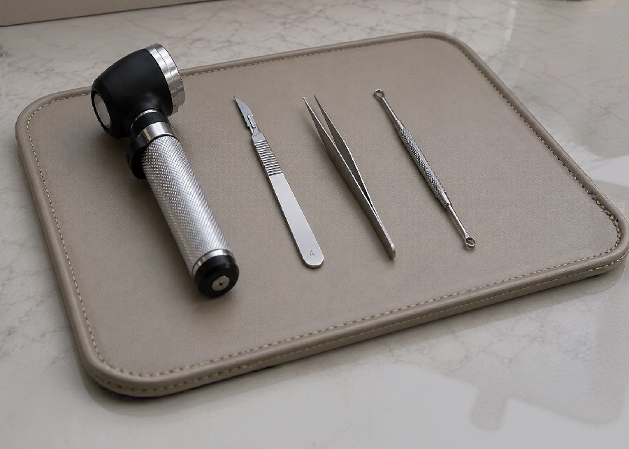
A dermatoscope, scalpel, forceps, and curette arranged on a soft leather tray

03, Surgical

Skin cancer & surgical care.

Early detection changes everything. Screening, biopsy, and years of protective follow-up.

Your visit

Straight answers, at every step.

Before

Note anything that has changed, bled, or refused to heal, and any history of skin cancer in your family. Come without makeup and with nail polish removed so every area can be seen.

During

A methodical head-to-toe exam with dermatoscopic review of anything of interest. What is being seen is described as it is found, and a biopsy can usually be done the same day.

After

Results explained directly, a plan you understand before you agree to it, and a return date set so nothing depends on remembering to call.

The care

End to end,
start to steady.

From the first exam onward you keep the same provider, the same chart, and the same eye watching for change. Open any card to read how each part of that care is evaluated, performed, and followed.

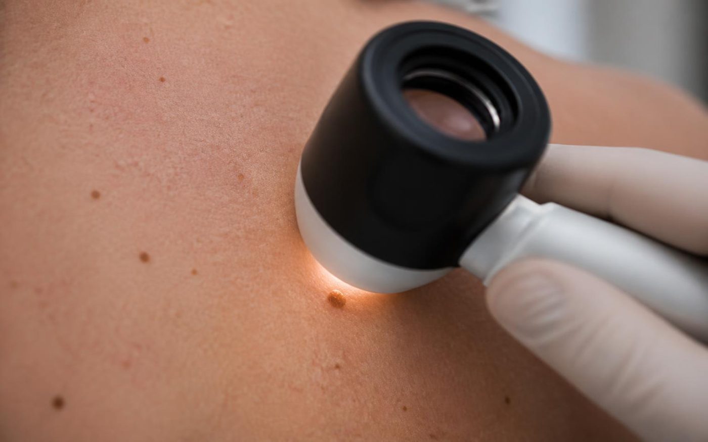
Dermatoscope held against the upper back to examine a small mole during a full-body skin exam


Full-Body Skin Exams

A careful head-to-toe evaluation with dermatoscopic imaging, documented so that any change can be recognized early.

Overview
A full-body skin exam is a systematic look at the skin from the scalp to the soles, including areas that are difficult to see on your own. A dermatoscope allows the structures beneath the surface of a mole or lesion to be examined in detail, which makes it possible to distinguish ordinary spots from those that deserve closer attention.
When it is recommended
An annual exam is reasonable for most adults, and more frequent visits are appropriate for anyone with a personal or family history of skin cancer, many or atypical moles, significant sun exposure or tanning history, fair skin that burns easily, or a history of immunosuppression.
How it is performed
You will change into a gown and the skin is reviewed in an orderly sequence in good lighting. Lesions of interest are examined with the dermatoscope and photographed when it is useful to compare them at a later visit. The exam is unhurried, and you are welcome to point out anything you have been watching.
What to expect during your visit
The exam itself usually takes fifteen to twenty minutes. Findings are explained as they are noted rather than saved for the end, so you understand what is being seen. Most visits conclude with reassurance and a date to return.
Follow-up
If a lesion is uncertain but not concerning enough to sample, a short-interval recheck is often the best next step. Anything suspicious is discussed at the same visit, and a biopsy can frequently be performed the same day.
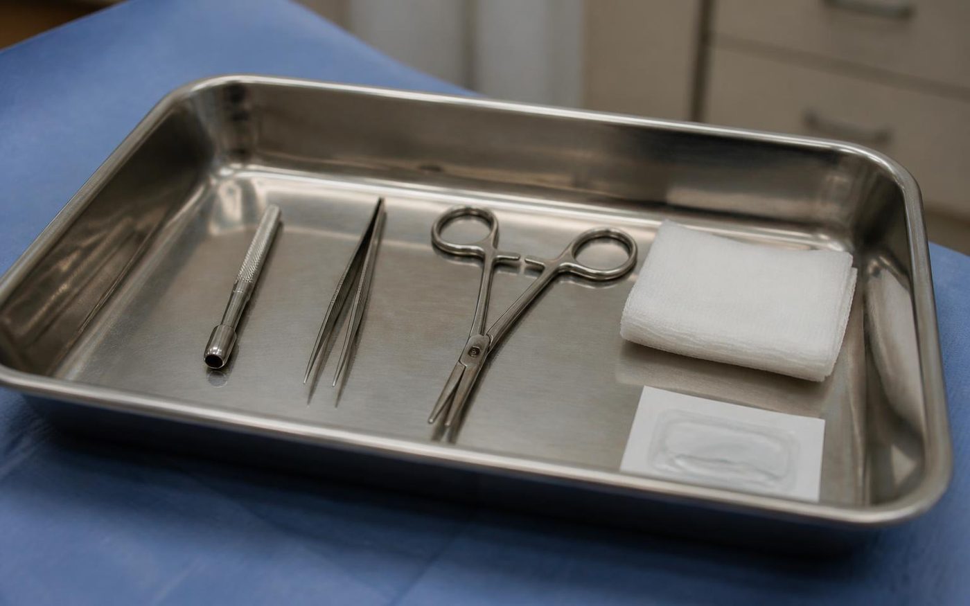
Sterile tray with a punch biopsy tool, forceps, needle driver, suture and gauze prepared for a skin biopsy


Biopsies

Same-visit sampling of a suspicious lesion, performed with local anesthesia and a light, precise technique.

Overview
A biopsy removes a small sample of skin so that it can be examined under a microscope. It is the only way to confirm what a lesion is with certainty, and it is a brief, routine office procedure rather than a surgical event.
When it is recommended
A biopsy is considered when a lesion has changed in size, shape, or color, when it bleeds or will not heal, when a mole looks different from your others, or when a rash has not responded to treatment and the diagnosis remains unclear.
How it is performed
The area is cleaned and numbed with local anesthetic. Depending on the lesion, the sample may be taken from the surface layers or through the full thickness of the skin, and a small suture is placed when needed. The sample is sent to a dermatopathology laboratory for review.
What to expect during your visit
You will feel a brief sting as the anesthetic is placed, and little else afterward. The procedure generally takes a few minutes, and you can return to your usual day. Written wound care instructions are provided before you leave.
Recovery and follow-up
The site is kept clean and covered while it heals, usually over one to two weeks, and any sutures are removed at a short follow-up. Results are typically available within about a week and are reviewed with you directly, along with what, if anything, comes next.
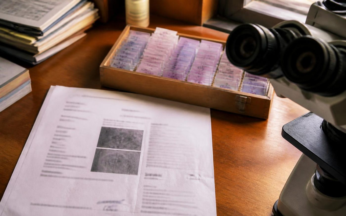
Dermatopathology slides and a pathology report beside a microscope during diagnosis review


Diagnosis & Evaluation

A clear conversation about results, what they mean, and every reasonable option in front of you.

Overview
Receiving a diagnosis is often the most difficult part of the process, and it is where clarity matters most. Skin cancers differ considerably in how they behave. Basal cell and squamous cell carcinomas are common and highly treatable, while melanoma requires prompt, more detailed planning. Understanding which one is present shapes everything that follows.
When evaluation is recommended
Evaluation follows any biopsy result that shows atypical or cancerous cells, and is also appropriate for a second opinion, for a diagnosis made elsewhere, or when you would like the findings explained more fully before deciding on treatment.
How it is approached
The pathology report is reviewed with you in plain language, including the type of lesion, its depth or margins, and what those details mean practically. Location, cosmetic considerations, your general health, and your preferences are all weighed before a recommendation is made.
What to expect during your visit
Time is set aside for questions, and you are encouraged to bring someone with you. You will leave understanding the diagnosis, the options, and the reasoning behind the recommendation rather than a single instruction.
Follow-up
Once a plan is chosen, the necessary appointments and referrals are arranged for you, and the practice remains your point of contact throughout.
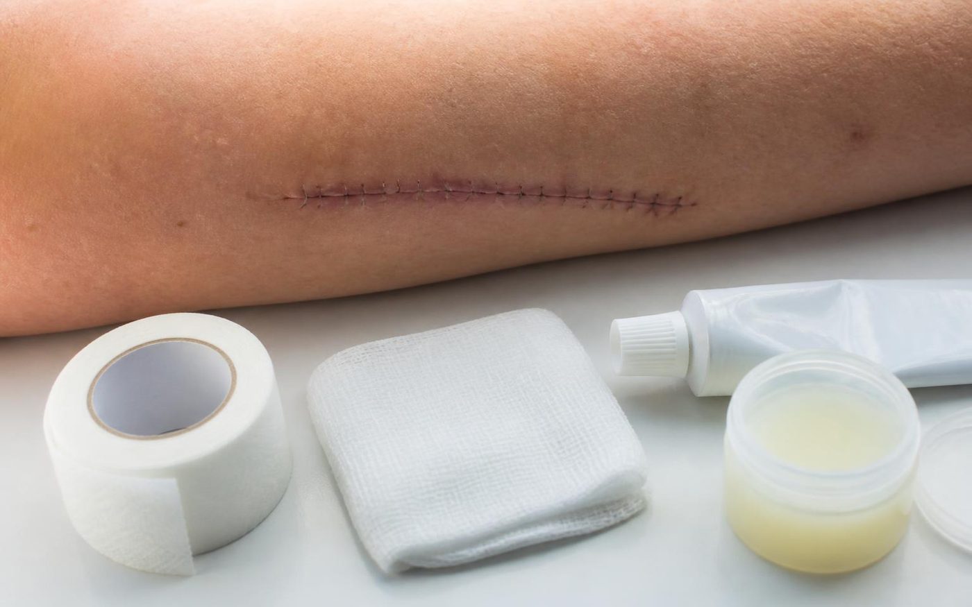
Healing sutured excision site on a forearm with tape, gauze, ointment and scar gel for aftercare


Pre & Post-Surgical Care

Preparation before a procedure, and attentive support through healing and scar maturation afterward.

Overview
The result of a procedure depends a great deal on what happens around it. Careful preparation reduces complications, and consistent aftercare influences how a scar settles over the months that follow.
When it applies
This care accompanies any biopsy or excision, whether performed here or with a surgical colleague, and is also appropriate if you are healing from a procedure done elsewhere and would like guidance.
How it is approached
Before the procedure, medications, supplements, and any conditions that affect healing are reviewed, and the day itself is explained so there are no surprises. Afterward, wound care is demonstrated rather than only described, and the site is rechecked at intervals appropriate to its location.
What to expect
Some swelling, bruising, and tenderness are ordinary in the first days. You will know what is expected, what is not, and exactly when to call. Written instructions accompany every procedure.
Recovery and follow-up
Scars remodel for a year or longer. Silicone, massage, sun protection, and in selected cases resurfacing or other treatment can improve the final appearance, and these are considered once the site has fully closed.
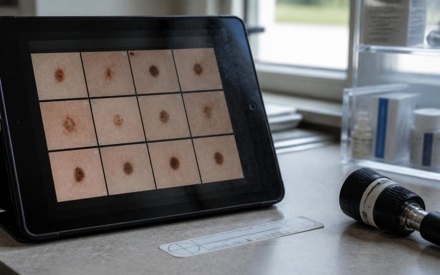
Mole mapping photographs displayed on a tablet with a measuring ruler and dermatoscope for ongoing surveillance


Long-Term Monitoring

Ongoing surveillance and prevention, because the most protective skin cancer care is the kind that continues.

Overview
Skin cancer risk does not end with treatment. Regular surveillance allows new or recurrent lesions to be found while they are small and simple to address, and it builds a record of your skin over time that makes change easier to recognize.
When it is recommended
Ongoing monitoring is advised after any skin cancer diagnosis, for those with numerous or atypical moles, for a strong family history, and for anyone with substantial past sun exposure or a history of immunosuppression.
How it is approached
Visits are scheduled at an interval matched to your risk, commonly every three, six, or twelve months. Prior photographs and notes are compared at each visit, and self-examination is reviewed so you know what to watch for between appointments.
What to expect during your visit
These are generally short, straightforward visits. Most end with reassurance, and when something does need attention it is usually caught at a stage where treatment is minor.
Follow-up
Intervals are adjusted as your risk changes, and anything new can be seen sooner. You are not asked to wait for the next scheduled visit if something concerns you.

Between visits

What is worth a closer look.

Most spots are harmless. These are simply the patterns worth mentioning at your next visit, or sooner if something is changing quickly.

Asymmetry

One half of a mole does not mirror the other.

Border

Edges that are uneven, notched, or fade into the surrounding skin.

Color

More than one shade within a single spot, or a color that has deepened.

Diameter

Larger than a pencil eraser, though smaller lesions can matter too.

Evolving

Any change in size, shape, color, or sensation over weeks to months.

The outlier

A spot that simply looks unlike the rest of your moles, or one that itches, bleeds, or will not heal.

The number that matters

Nine of ten skin cancers are curable when caught early.

Our Red Hook treatment room

Prevention

Habits that
quietly add up.

Prevention is unremarkable by design. A few consistent habits, reviewed at each visit, do more over a lifetime than anything dramatic.

  • Broad-spectrum SPF 30 or higher, worn daily and reapplied outdoors
  • Shade and protective clothing during peak midday hours
  • No tanning beds, at any age
  • A monthly self-check, including the scalp, back, and feet
  • An annual professional exam, or more often if your history calls for it

Once a year

Schedule a skin exam.

Twenty minutes, once a year, and often the most protective appointment you will make.